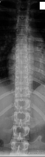
AP DORSAL
Anteroposterior Projection of Dorsal Spine • Evaluation of vertebrae T1 to T12
Exposure Factors
Medium exposure: Parameters for optimal visualization of thoracic vertebrae
Anatomical Structures Visible
Should be clearly observed:
- Dorsal vertebral bodies (T1 to T12)
- Pedicles
- Spinous processes
- Transverse processes
- Intervertebral disc spaces
Full field verification:
To ensure all dorsal vertebrae: See at least the last cervical vertebra (C7) and the first or second lumbar vertebra (L1/L2)
Cassette Size and Orientation
Longitudinal orientation to cover the entire thoracic spine
Patient Positioning
Alternative Position: Standing
If patient cannot lie down:
- Perform standing in wall bucky
- Same alignment and centering criteria
- Useful for patients with pain or limitation to lie down
Importance of Leg Flexion
"Legs should be bent so that the back lies completely in contact with the table"
Problems if legs are not flexed:
- Lumbar hyperlordosis that projects vertebrae incorrectly
- Incomplete contact of thoracic spine with table
- Distortion of intervertebral spaces
- Difficulty in evaluating vertebral alignment
Note: "Sometimes it's not done due to haste" - But it's essential for image quality
Central Ray Point
Location: Sixth thoracic vertebra
Angulation: Vertical and perpendicular to T6
Position: For patient in supine position
Standing alternative: Horizontal directed to T6
Optimal Image Characteristics
Vertebrae T1-T12
All included in field
Disc Spaces
Symmetric intervertebral
Symmetry
Spinous processes centered
Alignment
Straight-line spine
Transitions
C7 and L1/L2 visible
Pedicles
Symmetric and defined
Common Technical Challenges
Frequent problems in AP dorsal projection:
- Patient rotation causing spinous process asymmetry
- Hyperlordosis from not bending legs
- Incomplete field not including C7 or L1/L2
- Rib superposition on upper thoracic vertebrae
- Breathing during exposure causing blurring
- Incorrect centering cutting extreme vertebrae
Solution: Verify sagittal alignment, bend legs, instruct respiratory apnea
Special Considerations
Geriatric Patients
Marked kyphosis may require centering adjustment and possible angulation.
Obese Patients
Increase kV and mAs according to thickness adjustment table.
Bedridden Patients
Perform with cassette in direct, verify ray perpendicularity.
Patient Instructions
"Hold your breath during the exposure"
Maintain position without movement during radiographic exposure
1. "Bend your knees to support your back well"
2. "Stay completely straight"
3. "Do not turn your body"
4. "Take a deep breath and then hold your breath"
5. "Do not move during the exposure"